01 / The interface
Many modalities, one interface.
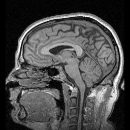
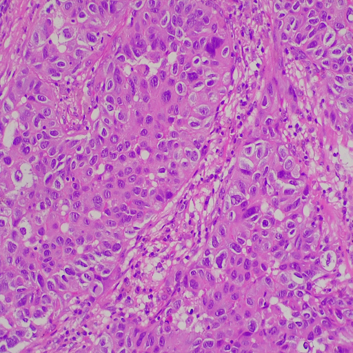
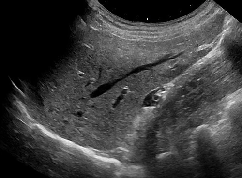
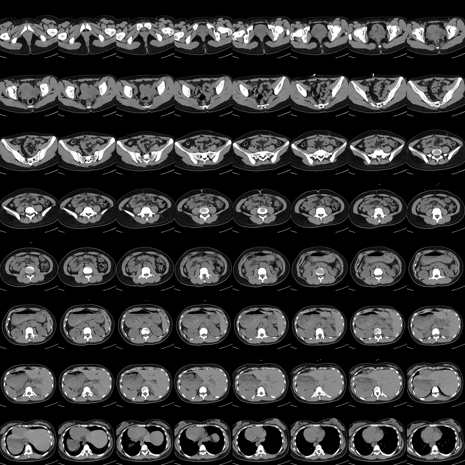
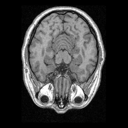
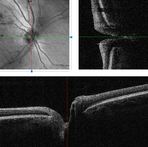
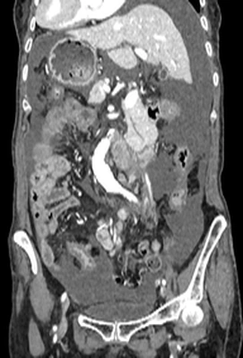
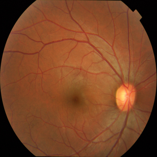
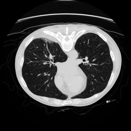
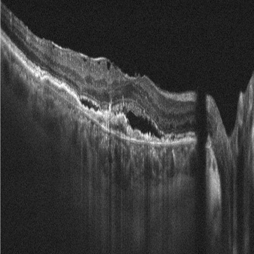
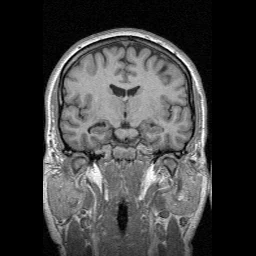
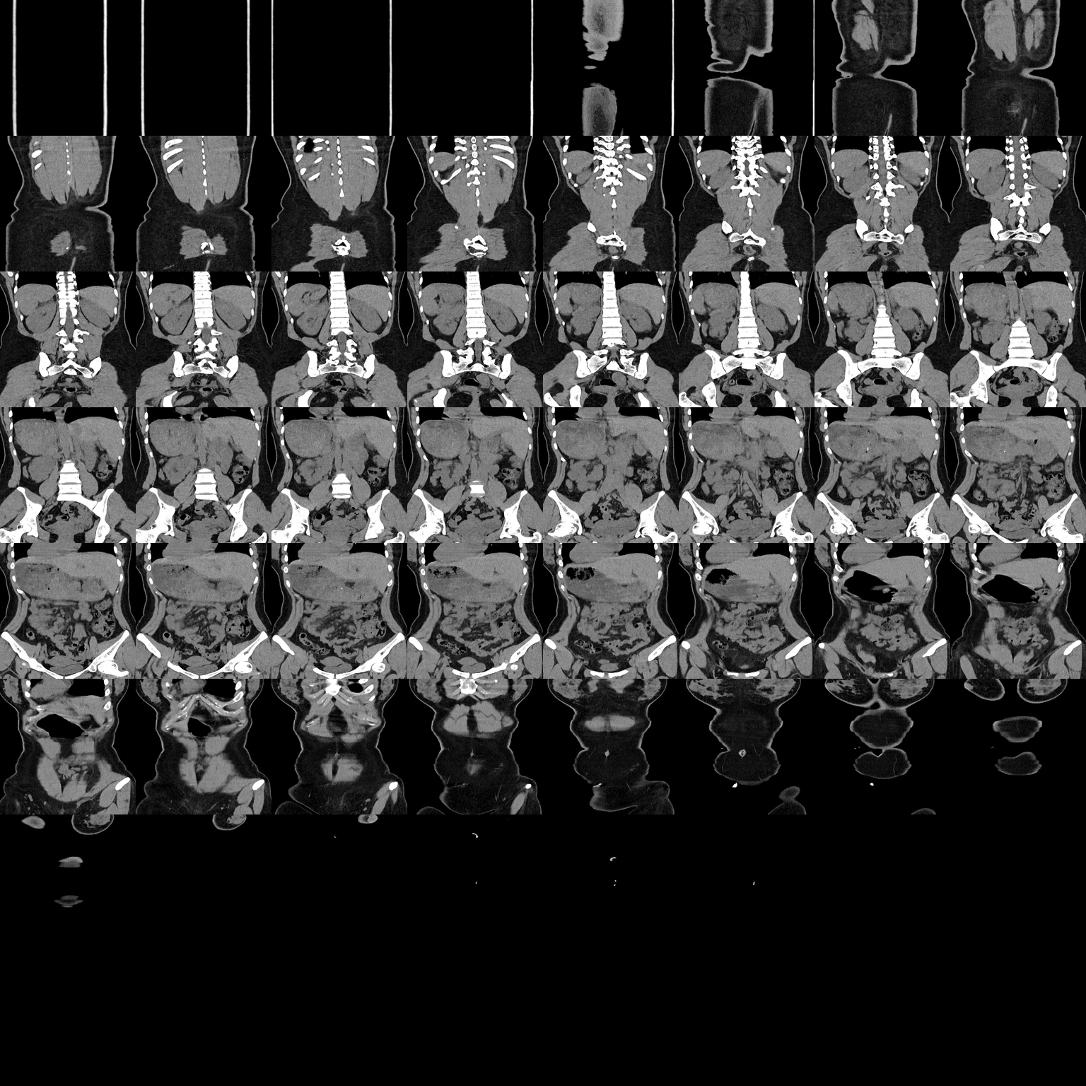
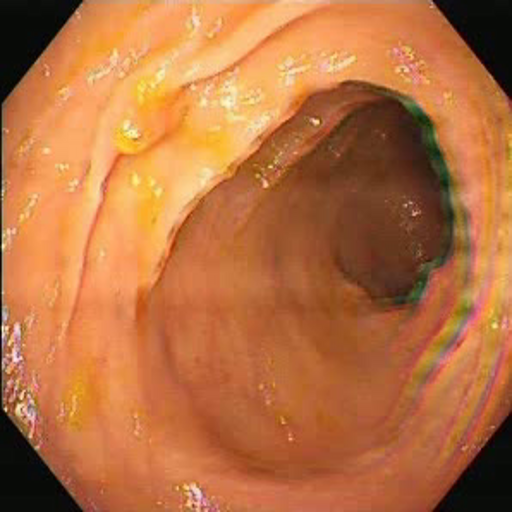
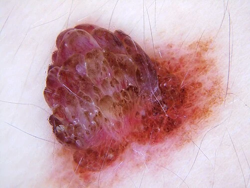
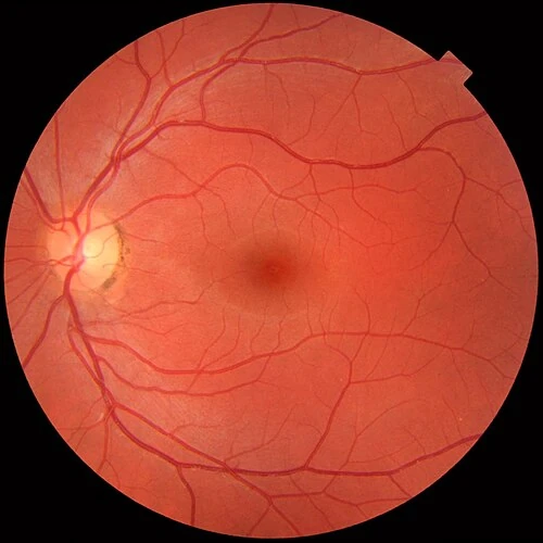
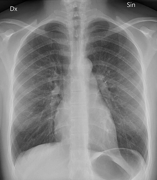
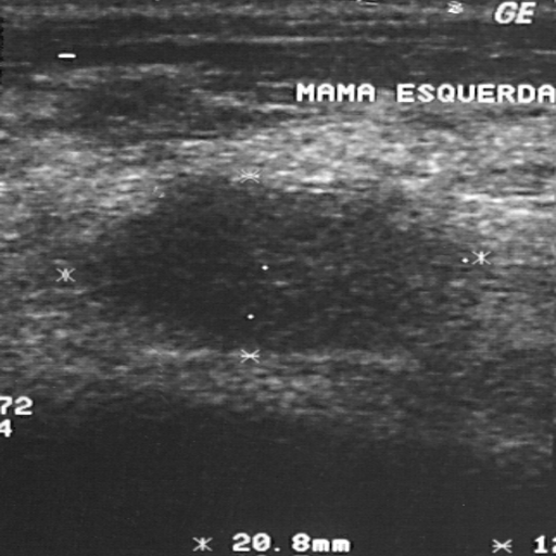
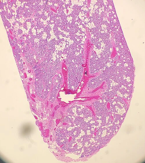
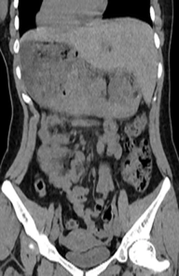
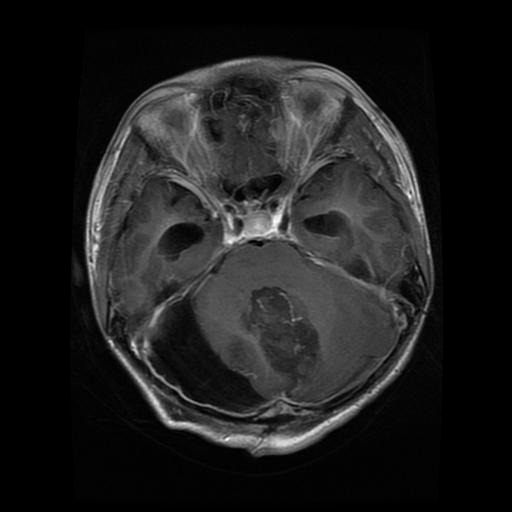
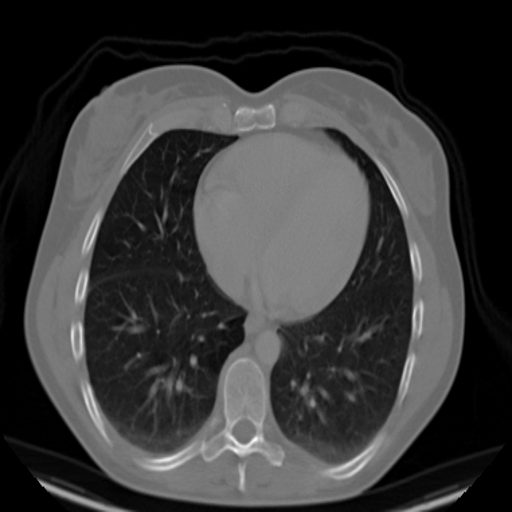
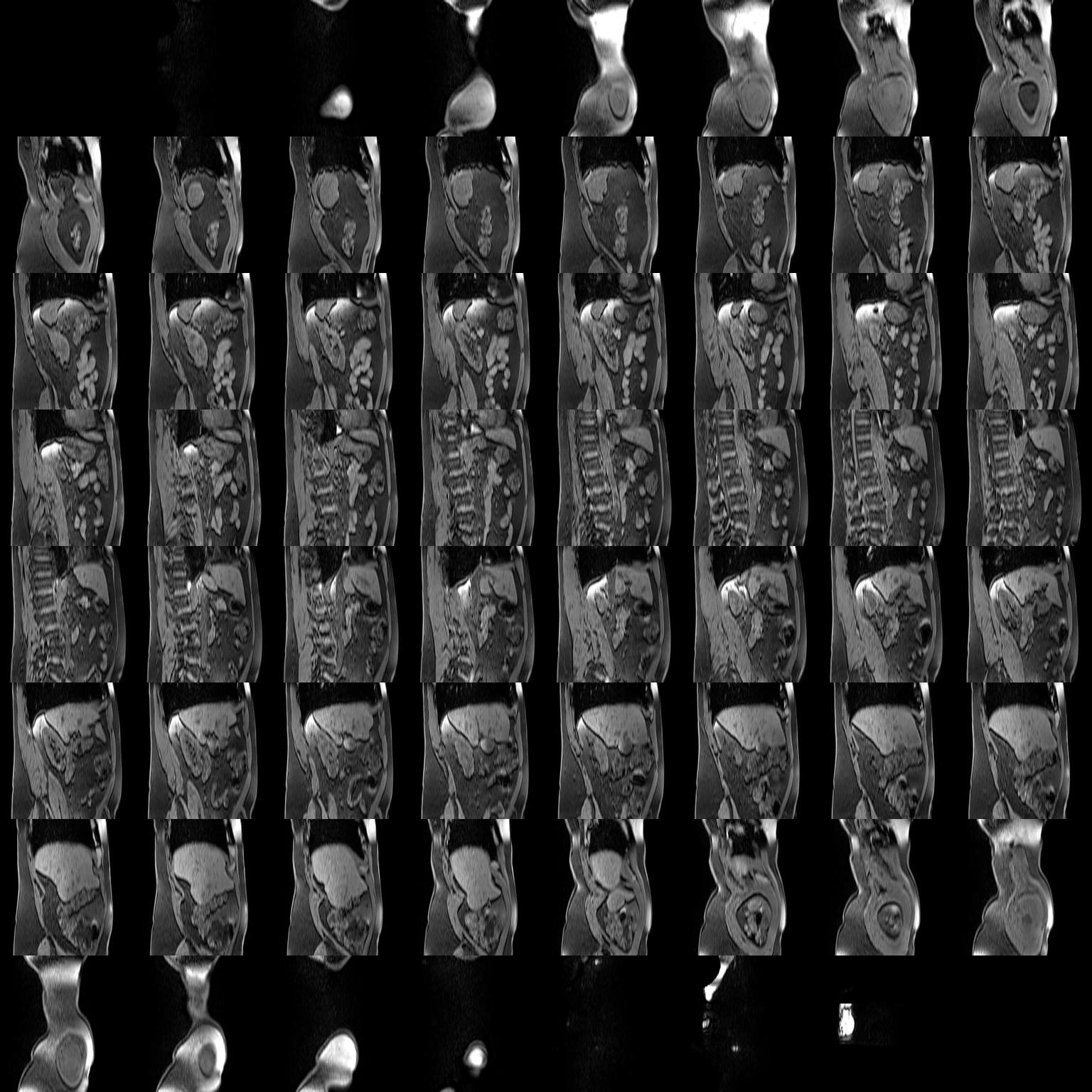
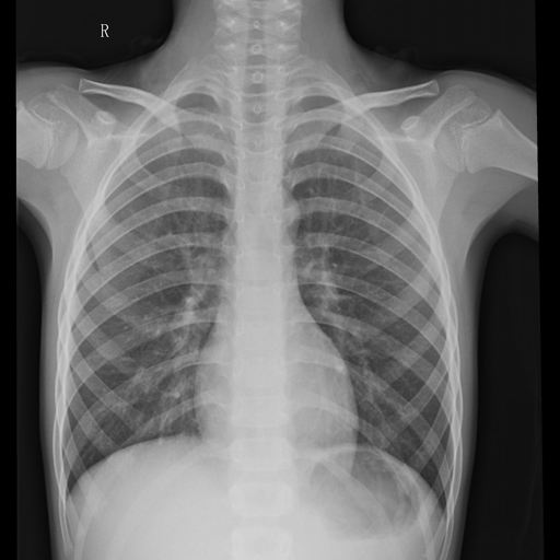
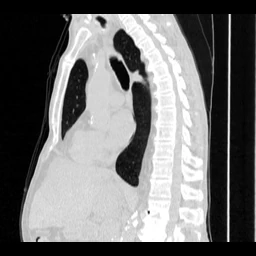
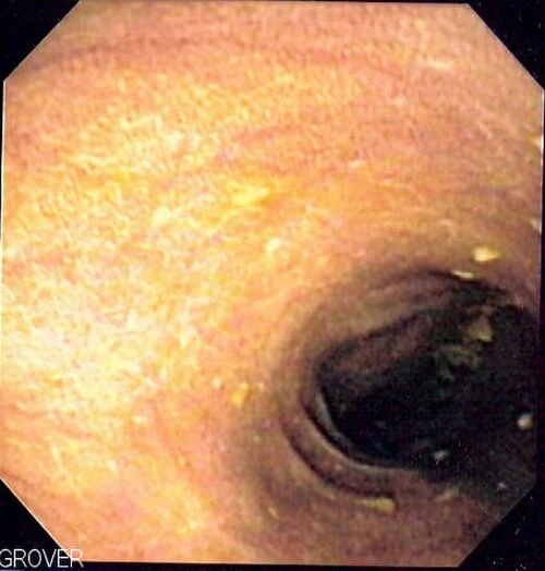
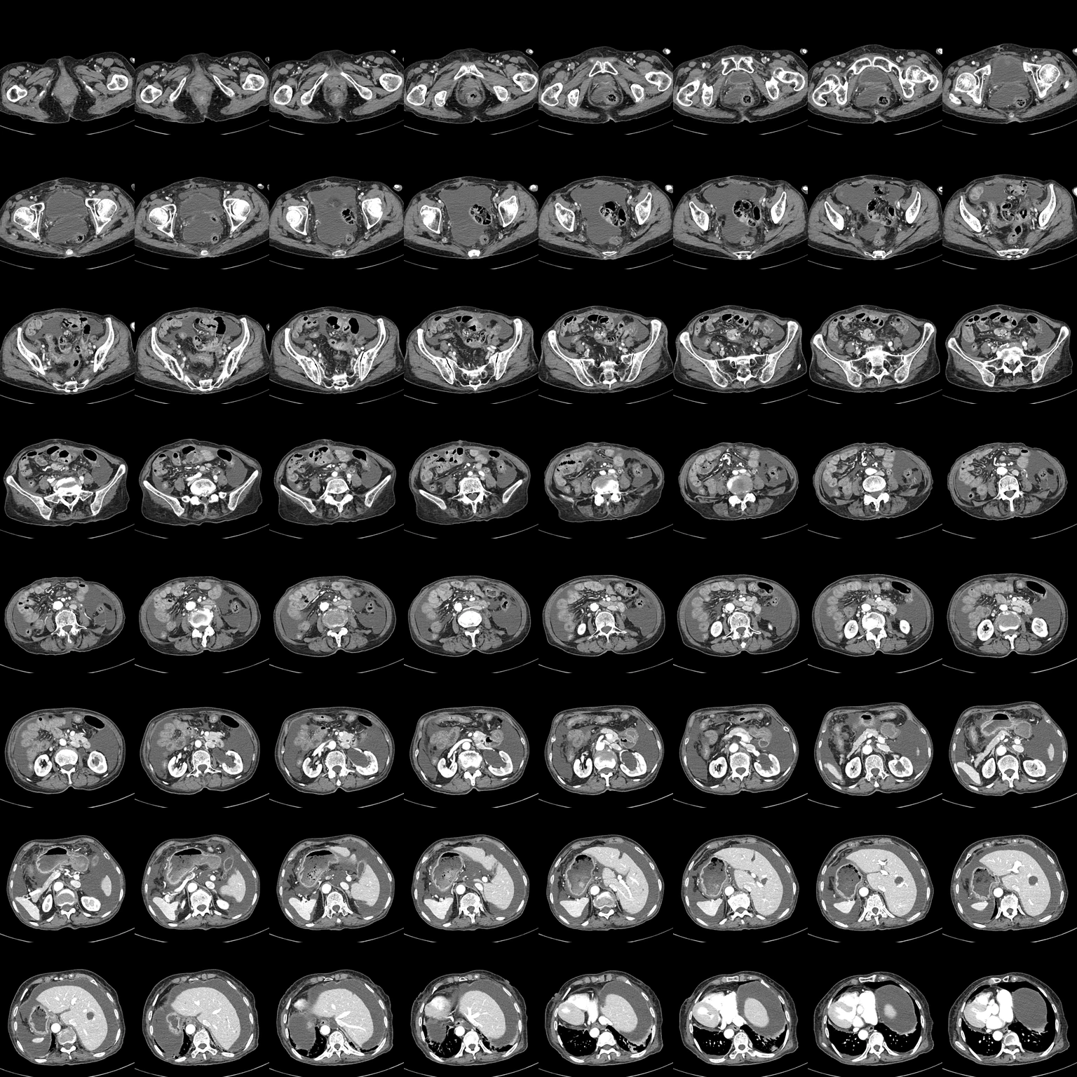
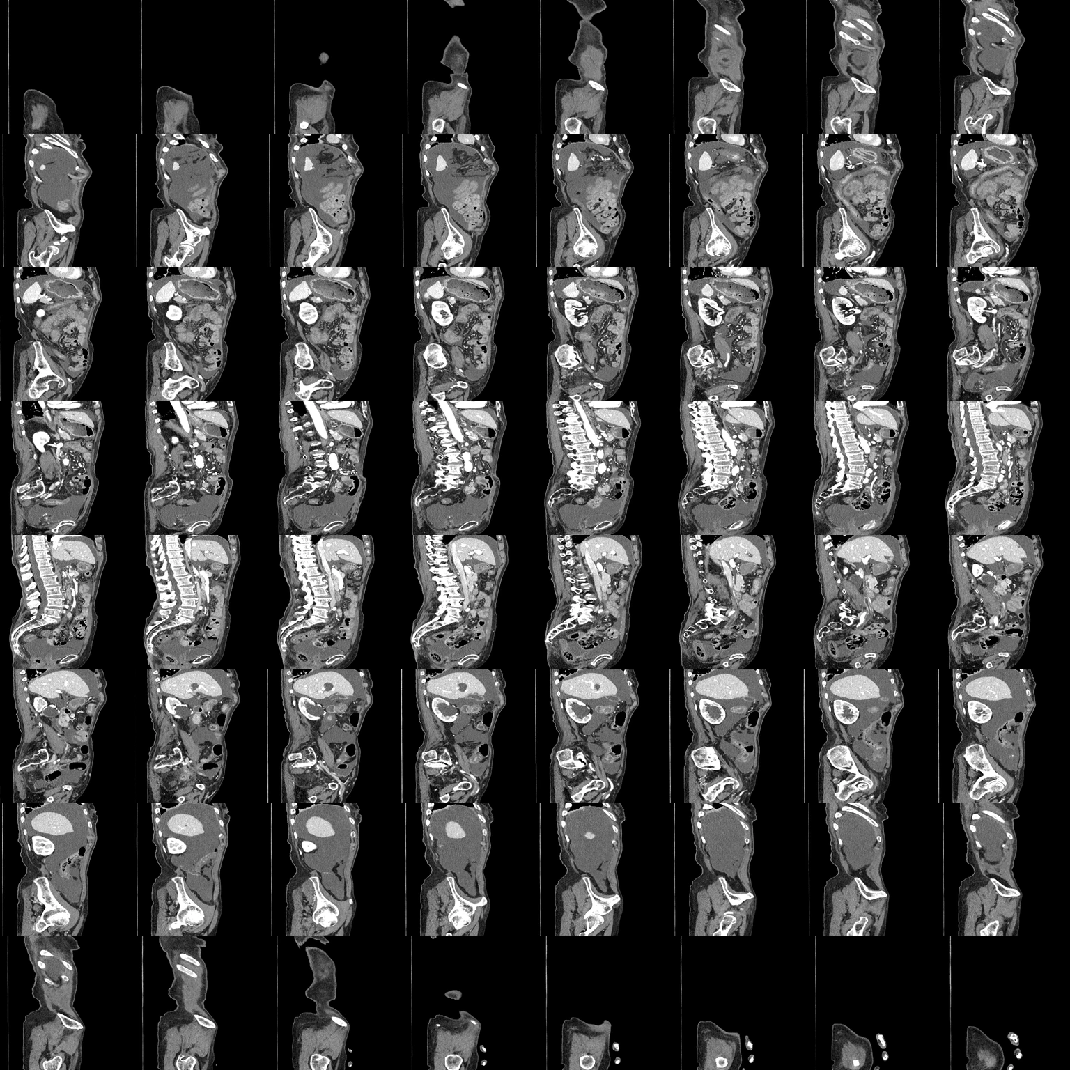
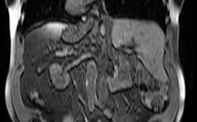
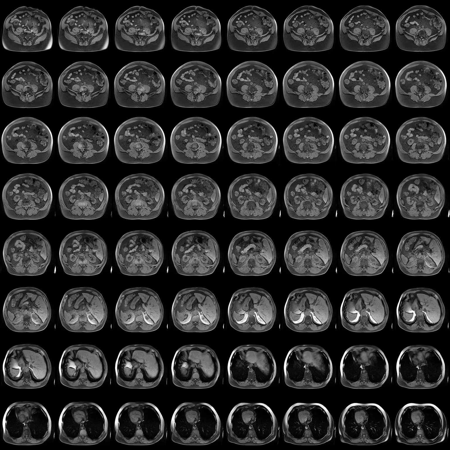
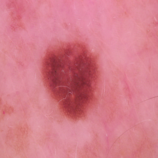
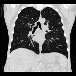
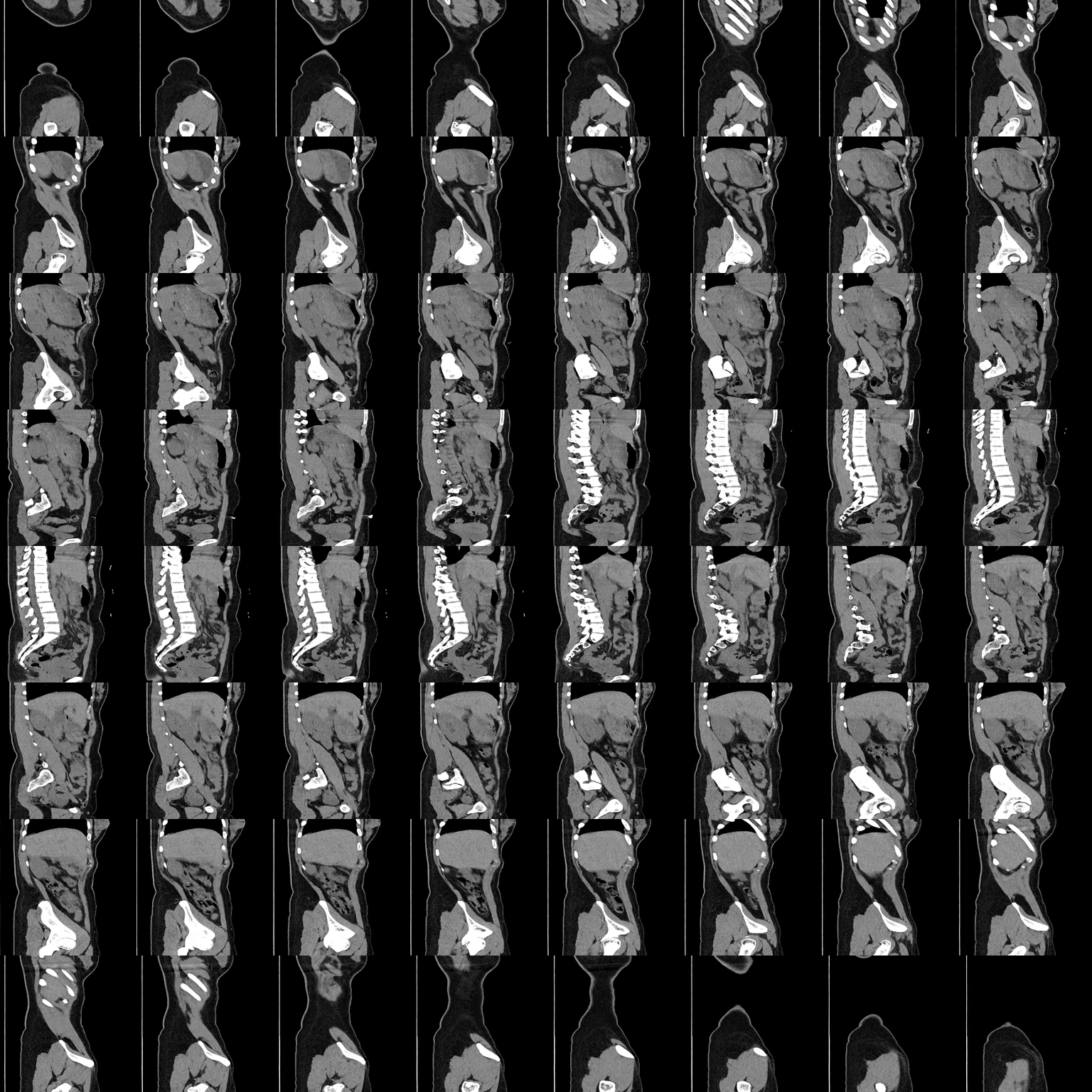
Medical imaging offers complementary views of the body. MedVTok encodes 2D images and 3D volumes across nine modalities into a common token interface that downstream models can reuse.
Diverse visual evidence
9 modalities · 2D images & 3D volumes
Unified medical visual tokenizer
MedVTok
Anatomical structure · Clinical semantics
Downstream tasks
Frozen tokenizer · task-specific models
02 / The approach
Many experts, one tokenizer.
Different medical imaging experts and foundation models capture complementary knowledge. MERA (Multi-Expert Representation Alignment) lets MedVTok learn from the specialist matched to each input. Shared-space alignment brings token and expert features together; alignment in the expert’s original space helps retain its distinct knowledge.
03 / The corpus
Training corpus.
We train this shared foundation on over 28 million 2D images and 70,000 3D volumes spanning nine imaging modalities.
| Modality | 28M+2D images | 70K+3D volumes |
|---|---|---|
| CT |
9.26M32.2%
|
35.3K50.3%
|
| Histopathology |
5.65M19.6%
|
— |
| MRI |
3.79M13.2%
|
34.9K49.7%
|
| Fundus |
3.05M10.6%
|
— |
| Endoscopy |
2.86M9.9%
|
— |
| X-ray |
1.59M5.5%
|
— |
| Dermoscopy |
1.26M4.4%
|
— |
| Ultrasound |
1.00M3.5%
|
— |
| OCT |
317.8K1.1%
|
— |
Rounded sample counts · Percentages within each dimension · — Not included
04 / Token fidelity
Preserve the visual evidence.
Reconstructing the input tests what the tokens preserve. MedVTok achieves the highest PSNR and SSIM among the compared tokenizers across nine 2D modalities and native 3D CT and MRI.
| Benchmarks | MedVTok | PUMIT | MedVAE | MedITok | AToken |
|---|---|---|---|---|---|
| 2D FundusJSIEC | 42.22 | 32.03 | 17.11 | 35.92 | 41.08 |
| 2D CTChestCTScan | 44.75 | 32.86 | 37.97 | 35.61 | 41.60 |
| 2D DermoscopyHAM10K | 40.39 | 27.05 | 22.42 | 33.81 | 39.00 |
| 2D EndoscopyPolypDiag | 41.42 | 28.73 | 16.83 | 32.42 | 39.69 |
| 2D MRIBRISC | 43.75 | 32.88 | 35.72 | 34.82 | 41.64 |
| 2D OCTOCTDL | 35.04 | 27.06 | 30.90 | 30.26 | 32.94 |
| 2D HistopathologyLungHist700 | 30.16 | 19.43 | 20.13 | 26.03 | 28.57 |
| 2D UltrasoundBUSBRA | 42.08 | 23.26 | 33.47 | 32.62 | 39.41 |
| 2D X-rayKermany | 41.83 | 26.69 | 37.04 | 34.70 | 40.02 |
| 3D MRIMRISegmenter | 31.77 | 29.96 | 25.89 | — | 31.46 |
| 3D CTAMOS-MM | 36.50 | 31.87 | 31.30 | — | 35.68 |
↑ Higher is better. Bold marks the best score in each row. — Not reported.
Benchmark data05 / Downstream utility
One frozen tokenizer, many tasks.
We then test how well the representation transfers. With MedVTok frozen, task-specific models use its features for classification, retrieval, segmentation, image synthesis, visual question answering, and report generation.
| Benchmarks | MedVTok | PUMIT | MedVAE | MedITok |
|---|---|---|---|---|
| ClassificationJSIEC · ChestCTScan · HAM10K · PolypDiag · BRISC · OCTDL · LungHist700 · BUSBRA · KermanyMean · Accuracy (%) ↑ | 80.23 | 52.41 | 51.32 | 73.55 |
| RetrievalJSIEC · ChestCTScan · HAM10K · PolypDiag · BRISC · OCTDL · LungHist700 · BUSBRA · KermanyMean · Recall@1 (%) ↑ | 79.18 | 52.53 | 51.30 | 74.43 |
| SegmentationMaplesDR · MosMedDataPlus · HAM10K · PolypDB · BRISC · PanNuke · BUSBRA · VinDr-RibCXRMean · Dice (%) ↑ | 76.07 | 53.63 | 38.86 | 71.99 |
| Image synthesisGMAI-VL-5.5MMean · FID ↓ | 101.73 | 227.51 | 172.08 | 109.76 |
| Visual question answeringFundus-MMBench · MM-Skin · Kvasir-VQA-x1 · PathVQA · SLAKE · GMAI-MMBenchMean · Accuracy (%) ↑ | 46.48 | 41.94 | 39.93 | 45.39 |
| Report generationAMOS-MMMean · GREEN ↑ | 0.413 | 0.250 | 0.230 | 0.386 |
Paper-reported scores with the tokenizer frozen. ↑ Higher is better; ↓ lower is better. Bold marks the best score in each row.
Benchmark data06 / Looking ahead
Toward a health world model.
MedVTok is a starting point for a broader goal: a coherent representation of a person’s health across modalities and over time.
We aim to connect medical images with clinical records and everyday health signals in a health world model that evolves as new observations arrive, supporting both patient care and lifelong well-being.
The team
MedVTok
A General-Purpose Medical Visual Tokenizer
Author affiliations
@inproceedings{ma2026medvtok,
  title  = {MedVTok: A General-Purpose Medical Visual Tokenizer},
  author = {Ma, Chenglong and Ji, Yuanfeng and Ning, Junzhi and Liu, Jiyao and Wei, Yujie and Rui, Shaohao and Zhang, Chaoyang and Li, Wenjie and Tang, Wenhao and Tang, Cheng and Li, Wei and Gao, Shujian and Zhang, Yujie and Chen, Ying and Li, Zilong and Ge, Zongyuan and Song, Diping and Ye, Jin and Hu, Ming and He, Junjun and Shan, Hongming},
  booktitle = {Advances in Neural Information Processing Systems (NeurIPS)},
  year   = {2026}
}
Acknowledgments
Illustrative image credits
The hero combines illustrative modality images and matched evaluation examples. CT and MRI orthogonal views come from 3D Slicer sample data. Reconstruction galleries use matched evaluation outputs. Synthesis, VQA and report examples are adapted from the paper.
- CT3D Slicer / Brigham and Women’s Hospital · 3D Slicer license
- HistopathologyMikael Häggström, M.D. · CC0 1.0
- MRI3D Slicer / Brigham and Women’s Hospital · 3D Slicer license
- FundusMikael Häggström · CC0 1.0
- EndoscopySamir at English Wikipedia · CC BY-SA 3.0
- X-rayMikael Häggström · CC0 1.0
- DermoscopyRaimundo Pastor · CC BY 4.0
- UltrasoundMikael Häggström · CC0 1.0
- OCTWies6014 · CC BY-SA 4.0